Breast Cancer

Breast Cancer

Being diagnosed with breast cancer is very frightening. Understanding the different types of cancer, is the first step in understanding treatment options. The discussion of breast cancer is multidisciplinary. The focus of treatment options and, insight into many aspects of breast cancer treatment is paramount to making informed decisions. These treatments may include surgery, radiation therapy, chemotherapy, hormonal blockade, and genetic testing.
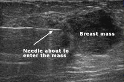
  • Invasive Cancers

    Invasive cancers are cancers {abnormal cells} that can spread to lymph nodes or other organs.  These types of cancers are seen under the microscope as having broken through the ‘basement membrane’ of the breast duct or lobule.  The ‘basement membrane’ is sort of a barrier to the blood vessels and lymphatics.  Once cancers cells have invaded beyond the ‘basement membrane’ they have the potential to spread to other organs (lymph nodes, liver, lung, etc.)


    There are different types of invasive breast cancer:

    • Infiltrating (invasive) ductal (75%)
    • Infiltrating (invasive) lobular (10%)
    • Mixed mammary (both ductal and lobular features (10%))
    • Special Subtypes: Colloid (1-2%), Tubular (1-2%) & Metaplastic (1-2%)
    • Inflammatory Breast Cancer (1-6%)
  • Non-Invasive Cancers

    Noninvasive cancers, are cancers {abnormal cells} that have not broken through the ‘basement membrane’. These cancers are also referred to as in situ or intraductal cancers.  In general, these types of cancers do not have the capacity to spread.  The incidence of this very early stage, non-invasive disease has expanded dramatically in recent years with more widespread mammographic screening.

  • In Situ Carcinoma

    As a consequence of population-based screening and the increased use of surveillance mammography, ductal carcinoma in situ (DCIS) now accounts for 20% of newly diagnosed breast cancers.   Lobular carcinoma in situ (LCIS) is not even considered cancer and is merely a ‘high risk lesion’.

  • Lobular Carcinoma In Situ (LCIS)

    Now also referred to as LIN – lobular intraepithelial neoplasia is a risk factor for subsequent invasive carcinoma in either breast with published relative risks of 8-10 times ‘average’.  So, not exactly ‘breast cancer’.  Treatment options of LCIS is that of all high-risk patients, thus complete excision, clear margins and radiation are not indicated.


    Pleomorphic LCIS – this is the exception; as this entity has features and behavior more like DCIS (ductal carcinoma in situ) but is still not considered or treated as cancer.  Excision to clear margins is a consideration and endocrine (hormonal) therapy is more strongly advocated.


    When to excise?  The greatest controversy surrounding LIN/LCIS is, when to surgically excise, when this diagnosis is made by core biopsy.  Multiple studies from different institutions have yielded upgrade/upstage rates (typically to DCIS) of 11-35%.  So, multiple factors go into making the decision whether surgical intervention is necessary.

  • Ductal Carcinoma In Situ (DCIS)

    The incidence of ductal carcinoma in situ has been steadily increasing with the widespread use of screening mammography and now accounts for approximately 20-25% of all breast malignancies.  Unlike invasive cancers, the size of DCIS does not have any impact on staging or outcomes.  The risk implied by having this diagnosis is that of recurrence of disease.  Patients with DCIS who recur will recur with invasive cancer in about 50% of cases.  So, the goal of treatment is to reduce this risk.


    Surgical options are like those for invasive cancer, including mastectomy and lumpectomy with radiation.

    • Local regional recurrence rates are similar but not zero for any surgical treatment.  Even women treated with mastectomy have a 0.8-3.3% risk of local recurrence.
    • The need for clear margins (lumpectomy) is also similar to invasive cancer patients, prior to radiation therapy.
    • There is no indication for chemotherapy in women with pure DCIS.

    There has been growing evidence to support the fact that not all women with DCIS need to be treated in the same fashion.  There are likely subsets of the disease whereby certain treatments may offer little if any benefit.  Being able to accurately predict which subset requires which treatment is currently under investigation.

  • Male Breast Cancer


    According to cancer researchers, it’s estimated that in 2014 there will be 2,360 new cases of male breast cancer in the U.S. This is in contrast to 232,670 new breast cancer cases projected for women.


    Wait, so how is male breast cancer possible? Male breast cancer may seem strange to think about, but it’s easier to grasp when you consider something we all have in common: breast tissue. Like women, men have breast tissue—it may be less “fatty” because they have different hormones that stimulate less breast growth, and its incapable of producing milk—but this tissue is still vulnerable to cancer.


    Here’s how male breast cancer forms: First, cells in the breast tissue change and they stop carrying out their normal cell function. Over time, the healthy cells will naturally die off and the body will replace them with malignant cancer cells, which don’t die off like normal cells. These abnormal cells form together as a lump or “tumor,” and their damaging behavior can be lethal.


    Symptoms of Male Breast Cancer - Early detection is imperative for survival, but most males don’t think to check for breast cancer so it often goes unnoticed. Here are some common symptoms to look out for:


    •  A lump in breast tissue
    •  Pain or throbbing in nipple area
    •  An inverted nipple
    •  Fluid or blood discharge from the nipple
    •  Inflammation or sores of the nipple/areola
    •  Swelling of the lymph nodes under the arm

    Breast enlargement can occur in men, but it’s not always a sign of cancer. Known as “gynecomastia,” this condition causes a man’s breast to become quite large, usually because of a hormone imbalance. Some causes for this include certain medications, excessive alcohol consumption, severe weight gain, and marijuana use.


    Treatment of Male Breast Cancer - Treatment for breast cancer in men is often handled in the same way as women. In short, it depends on the stage of the cancer and how well the patient is doing. Most men with breast cancer have a mastectomy, which is a surgical procedure that involves removing the breast, the lining of the chest muscles, or portions of the underarm.


    If the cancer has spread or metastasized, then doctors may recommend chemotherapy, radiation therapy, targeted therapy, or hormone therapy. These treatment options are called “adjuvant therapies,” and they are used to kill cancer cells, stop cancer growth, or a combination of both.

Breast Biopsy

Breast biopsies are sometimes required to make a definitive diagnosis. If the lesion of concern is too small to appreciate by ‘feel’, a needle localization may be necessary. Needle localization, is done in the Breast Imaging Department. A mammogram or ultrasound (sometimes an MRI) is used to identify the lesion of concern, and, then to guide a wire, placed next to (adjacent) to this area of concern. This localizing wire, will assist surgically, in identifying the area of concern.

Subareolar dissection is a procedure that is sometimes performed in women who have unexplained nipple discharge. This is a procedure, whereby, the affected (draining) duct is isolated and a portion removed. Similar to a breast biopsy, the procedure sometimes requires a preoperative localization. Although most nipple discharge is from a benign (noncancerous) etiology, biopsy/subareolar dissection is at times required.

  • Tissue Biopsy

    Once an abnormality is identified in the breast, the next step is obtaining a tissue diagnosis.  A tissue diagnosis will allow a treatment plan to formulate.  In the past, all biopsies of the breast were done surgically.  Today, the standard has changed.


    When it comes to biopsy of the breast, there is consensus. The American Society of Breast Surgeons:


    Whenever possible, diagnosis of breast lesions should be performed with a needle, rather than a surgical procedure. When a lesion is seen with ultrasound, this modality should be used for image-guidance. Image-guidance is an extremely useful adjunct in the performance of percutaneous biopsy of palpable breast lesions. Image-guidance confirms the proper placement of the biopsy device into the lesion (when core needle device is used), or immediately below the lesion (when vacuum-assisted device is used). Performing percutaneous breast biopsy procedures without the use of image-guidance may lead to false negative results since the biopsy device cannot be confirmed to be in the proper position to obtain tissue from the suspect mass. In most, if not all instances of image-guided biopsy of palpable breast lesions, ultrasound is the preferred image-guidance modality.

  • Ultrasound Guided Core Biopsy

    When a lesion is visible by ultrasound, this modality is used to localize the lesion, and, guide biopsy.  Ultrasound guided procedures are well tolerated.  This procedure can be done in the office, typically, on the same day as the office visit.

  • Stereotactic Core Biopsy

    A stereotactic core biopsy, is a technique whereby a lesion that is visualized mammographically, can undergo biopsy, using mammographically (stereotactic) guided biopsy techniques.  This again, follows the recommendation of using a needle to make the diagnosis of a breast lesion.  Stereotactic core biopsies are performed when a lesion is visible by mammogram only.

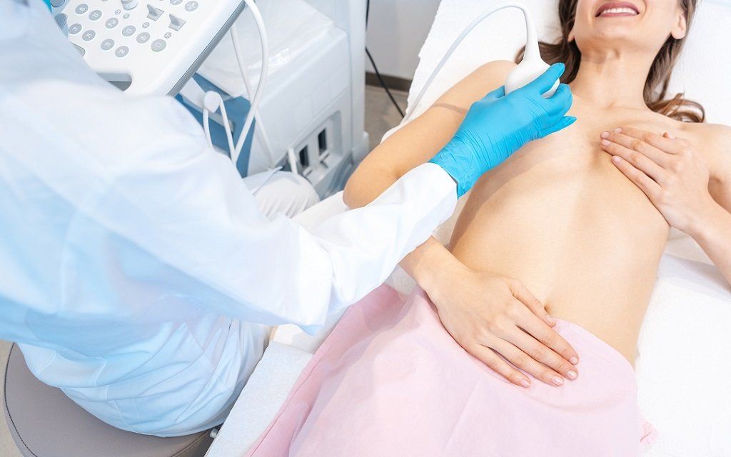
Imaging and Diagnostic Procedures

There are multiple imaging modalities utilized in diagnosing disease of the breast: breast ultrasound, breast MRI, tomosynthesis or 3D mammography, image guided core needle biopsies (ultrasound, MR, and, stereotactic guided), as well as, less conventional studies like thermascan. The goal of diagnostic imaging and biopsy is to utilize the least invasive technique to make a definitive diagnosis. The most critical part of making a diagnosis, is determining the appropriate testing needed. Just because there ‘is a test’ doesn’t mean it is necessary. The right test for the right patient is paramount to high quality care.

  • Mammography

    The “gold standard”.  Screening mammography remains the best method to screen the breast.  Typically, screening studies involve two views of each breast, and look for symmetry, masses, and calcifications.  Like all early detection strategies, screening mammography involves trade-offs.  Understanding these trade-offs is imperative in joint decision making as to when and in whom screening mammography should be performed.

  • Automated Breast Ultrasound (ABUS)

    Automated breast ultrasound (ABUS) is used as an adjunct to mammography for screening breast cancers in asymptomatic women with dense breasts. It is an effective screening modality with diagnostic accuracy comparable to that of handheld ultrasound (HHUS). Typically, ultrasound is not a screening test, however, there are ongoing Clinical Trials evaluating it’s use in this context. Ultrasound guided biopsies/aspirations are done, typically on the same day as an office visit, using local anesthetic. Expediting diagnosis in this manner, hopefully can reduce some of the anxiety caused by having an abnormal study. In addition, those women with a new mass/lump, may have a same day ultrasound, in order to better direct the next step in diagnosis and care. 

  • Breast MRI

    Magnetic resonance imaging (MRI) is a noninvasive medical test.  MRI uses a powerful magnetic field, radio frequency pulses and a computer to produce a detailed picture of the breast. MRI does not use ionizing radiation (x-rays).  Breast MRI requires the injection of a ‘dye’ gadolinium intravenously to help generate the multiple pictures of the breast.  What is a little peculiar, is that when the test is being performed, women lie prone (face down).  Both breasts are imaged.  The test typically takes 30-45 minutes.

Share by: